
Biopsy Test in Pokhara — Ultrasound-Guided Core (Trucut) Biopsy
A biopsy test in Pokhara at KDRC is a procedure in which a small tissue sample is taken from a lump or abnormal area, usually with a core (trucut) needle under ultrasound guidance and local anaesthetic, and sent to a pathology laboratory. It shows whether the area is benign, infected or cancerous and guides treatment.
Also called: biopsy test, tissue biopsy, ultrasound guided biopsy, usg guided biopsy, trucut biopsy, tru cut biopsy
 BiopsyKDRC · Pokhara
- Duration
- Most ultrasound-guided needle biopsies are completed within about an…
- Reported by
- Consultant radiologists
- Location
- Amarsingh Chowk, Pokhara-10
- Book / price
- 061553538 · 9856077433
 Dr. Nawaraj Paudel · Consultant RadiologistMedically reviewed by Dr. Bishworaj Bastakoti · MD Radiology Last reviewed 12 Oct 2026Updated 12 Oct 2026
- A biopsy takes a small tissue sample so a pathologist can make a definite diagnosis.
- Ultrasound-guided core (trucut) biopsies at KDRC are performed by consultant radiologists under local anaesthetic.
- FNAC collects loose cells; a core biopsy collects tissue that shows structure.
- Tell us about blood thinners before your biopsy and follow any fasting advice.
- Prices are quoted individually; call 061-553538 or 9856077433 for the current cost.
On this page (14 sections)
- What is a biopsy?
- Why is a biopsy needed?
- Types of biopsy
- FNAC vs core (trucut) biopsy: which is right for you?
- Ultrasound-guided biopsy in Pokhara
- How to prepare for a biopsy
- What happens during an ultrasound-guided biopsy at KDRC?
- Is a biopsy painful? Risks and safety
- Biopsy aftercare and recovery
- Biopsy results: what happens at the pathology laboratory?
- Biopsy in special situations: pregnancy, children and blood thinners
- How much does a biopsy cost in Pokhara?
- Why choose KDRC for a biopsy in Pokhara?
- Booking a biopsy and getting to KDRC
What is a biopsy?
A biopsy test in Pokhara at Kaligandaki Diagnostic and Research Center (KDRC) is a procedure in which a small sample of tissue is taken from a lump or abnormal area, usually with a needle under ultrasound guidance, and sent to a pathology laboratory for examination under a microscope. The NHS defines a biopsy simply as taking a small sample of skin, tissue or cells from the body for testing.
A biopsy is often the step that turns "something seen on a scan" into a firm diagnosis. Imaging such as ultrasound or a CT scan can show the size, shape and position of a lump, but only a pathologist looking at the tissue can say exactly what it is. That answer guides treatment, whether it is reassurance, medicine, follow-up or surgery.
At KDRC, needle biopsies are performed by consultant radiologists under live HD ultrasound guidance and local anaesthetic. Many patients in Nepal simply call it "बायोप्सी" or a tissue test. Most go home a short while after the procedure.
Why is a biopsy needed?
A biopsy is needed when a doctor must know what an abnormal area is made of before deciding on treatment. Common reasons include a lump felt on examination, a suspicious finding on ultrasound, mammogram or CT, an enlarged lymph node that does not settle, or an earlier FNAC that was inconclusive.
- To tell whether a lump is benign (not cancer) or malignant.
- To identify infection or inflammation, such as tuberculosis in a lymph node.
- To find the exact type of a tumour, which affects treatment choices.
- To confirm a diagnosis suggested by imaging or blood tests.
A pathology report usually describes what the tissue looks like, names the diagnosis where possible, and may list the results of special stains. For tumours it may include the type and other features that help specialists choose treatment. Your doctor will explain what each part means for you.
Being asked to have a biopsy does not mean you have cancer. Many biopsies show benign conditions. MedlinePlus, for example, notes that abnormal lymph node results range from mild infections to serious disease, which is exactly why a tissue answer matters.
Types of biopsy
Biopsies differ mainly in how the sample is collected: with a fine needle, a core needle, an endoscope, or by surgery. The NHS describes these routes as cutting out all or part of an area, using a needle to collect cells or tissue, or using an endoscope. The right method depends on where the abnormal area is, how big it is and what question your doctor needs answered.
| Type | How the sample is taken | Typical uses | At KDRC |
|---|---|---|---|
| Fine needle aspiration (FNAC) | Very thin needle draws loose cells | Thyroid nodules, lymph nodes, cysts | Yes, see FNAC |
| Core needle (trucut) biopsy | Spring-loaded hollow needle removes tissue cores | Breast lumps, masses, lymph nodes | Yes, ultrasound-guided |
| Vacuum-assisted biopsy | Suction draws larger tissue samples through one needle | Some breast lesions | Ask our team |
| Endoscopic biopsy | Tiny forceps through an endoscope | Oesophagus, stomach, duodenum | Yes, during endoscopy |
| Excision or incision (surgical) biopsy | Surgeon removes all or part of a lump | Some lymph nodes, skin and soft-tissue lumps | Your surgeon arranges this |
The word "trucut" (from a brand name) is widely used in Nepal and India for a core needle biopsy. If your report or referral says "trucut biopsy", it means a core needle biopsy.
FNAC vs core (trucut) biopsy: which is right for you?
FNAC collects loose cells and is quick and gentle, while a core (trucut) biopsy collects thin cylinders of tissue that show how the cells are arranged. RadiologyInfo explains that in FNA a thin needle on a syringe withdraws fluid or clusters of cells, whereas a core needle uses a spring-loaded mechanism to fill the needle with tissue cores, repeated several times. Both samples go to a pathology laboratory.
| Feature | FNAC | Core (trucut) biopsy |
|---|---|---|
| Sample | Cells and fluid on slides | Tissue cores in preservative |
| Laboratory test | Cytology | Histopathology |
| Needle | Very fine | Larger, spring-loaded |
| Anaesthetic | Often optional | Local anaesthetic routinely |
| Information | Cell type | Cell type plus tissue structure; extra special stains possible |
| Recovery | Immediate | Brief; avoid strenuous activity for about 24 hours |
| Best when | A quick first answer is enough | A detailed or definitive diagnosis is needed |
Your doctor and radiologist make the final choice together. Sometimes both are used: an FNAC first, then a core biopsy if the cells are not conclusive.
Ultrasound-guided biopsy in Pokhara
An ultrasound-guided biopsy uses live ultrasound images to steer the needle precisely into the abnormal area while avoiding blood vessels and other structures. RadiologyInfo notes that image-guided biopsies use ultrasound, CT, fluoroscopy or MRI to place the needle, and that the abnormal area must be visible on imaging. Ultrasound has the advantages of showing the needle in real time and using no radiation.
At KDRC, our consultant radiologists scan the area first on HD ultrasound, often with colour Doppler to see blood vessels, then plan the shortest safe path. Common targets for ultrasound-guided biopsy include lumps in the breast, thyroid, neck lymph nodes and soft tissues. Whether a particular organ or deeper lesion can be sampled safely at KDRC depends on its size, depth and position, so please ask our team with your scan report in hand.
Breast biopsy in Pokhara
A breast biopsy is usually an ultrasound-guided core needle biopsy that samples a breast lump or a suspicious area seen on imaging. According to RadiologyInfo, the doctor typically takes three to six core samples, the procedure is usually completed within an hour, you stay awake with local anaesthetic, and no stitches are needed.
- Safety: RadiologyInfo reports bleeding or a haematoma in less than 1% of patients and infection needing antibiotics in less than 1 in 1,000.
- Marker clip: a tiny marker may be left at the biopsy site so it can be found again; RadiologyInfo notes markers are MRI compatible and do not set off metal detectors. Ask our team whether a marker is planned in your case.
- Aftercare: mild swelling and bruising are common; avoid strenuous activity for at least 24 hours.
Most breast lumps that are biopsied are not cancer. The pathologist's report, checked against the imaging, tells your doctor what the next step should be.
How to prepare for a biopsy
Preparation for a needle biopsy is usually simple, but blood thinners, fasting and transport need planning. RadiologyInfo advises telling your doctor about all medicines, including blood thinners and herbal supplements, allergies and any chance of pregnancy; doctors may ask you to stop blood thinners beforehand and sometimes to fast for about eight hours. Our staff will give you exact instructions for your biopsy when you book.
| Item | What to do | Why |
|---|---|---|
| Blood thinners | Tell us when booking; your prescribing doctor decides whether to pause them | Reduces bleeding risk |
| Food and drink | Eat normally unless told to fast; follow our instructions | Some guided or sedated biopsies need an empty stomach |
| Diabetes medicines | Ask about dose changes if fasting | Avoids low blood sugar |
| Allergies | Mention reactions to local anaesthetic, antiseptic or plaster | Allows safe alternatives |
| Pregnancy | Tell us if you are or may be pregnant | Affects planning and positioning |
| Documents | Referral, earlier scans, FNAC or biopsy reports | Helps target the right area |
| Companion | Bring an adult if sedation is planned | You should not travel home alone after sedation |
- Tell us about blood thinners and herbal supplements when booking
- Do not stop any medicine without your prescribing doctor's advice
- Follow any fasting instructions our staff give you
- Mention allergies, diabetes and pregnancy
- Bring your referral, earlier scans and reports
- Bring an adult companion if sedation is planned
- Wear comfortable clothing that gives easy access to the area
What happens during an ultrasound-guided biopsy at KDRC?
An ultrasound-guided biopsy at KDRC is an outpatient procedure under local anaesthetic, usually completed within an hour including preparation. The steps below describe a typical core needle biopsy.
- Check-in and consent. Staff confirm the site, medicines and allergies; the radiologist explains the procedure and its risks and takes your consent.
- Scan and mark. The radiologist scans the area and plans the needle path, sometimes marking the skin.
- Clean and numb. The skin is cleaned and local anaesthetic is injected, which stings for a few seconds.
- Take the samples. A tiny nick lets the needle pass. You may hear a click as the spring-loaded needle takes each core, and feel pressure but not sharp pain.
- Dressing. Pressure stops any bleeding and a small dressing is applied; stitches are not usually needed.
- Observation. You rest briefly before going home with aftercare advice.
- 1Check-in and consentStaff confirm the site, medicines and allergies; the radiologist explains the procedure and takes consent.
- 2Scan and planThe radiologist scans the area on HD ultrasound and plans a safe needle path.
- 3Clean and numbThe skin is cleaned and local anaesthetic is injected.
- 4Take the coresThe biopsy needle takes several tissue cores under live ultrasound view.
- 5Dress the sitePressure stops bleeding and a small dressing is applied.
- 6Rest and go homeAfter brief observation you leave with aftercare advice; samples go to the pathology laboratory.
Is a biopsy painful? Risks and safety
A needle biopsy is usually not painful because local anaesthetic numbs the area; most people feel a brief sting from the injection and then pressure. MedlinePlus describes the anaesthetic as a brief prick and mild stinging, with the site sore for a few days. Tell the team at once if you feel sharp pain, as more anaesthetic can be given.
- Common: soreness and bruising for a few days, eased by simple painkillers such as paracetamol, as the NHS notes.
- Uncommon: a haematoma (collection of blood) under the skin.
- Rare: infection, significant bleeding, or injury to nearby structures. RadiologyInfo reports infections needing antibiotics in fewer than 1 in 1,000 needle biopsies.
- Limitations: sometimes too little tissue is obtained and the biopsy must be repeated.
Contact your doctor or go to the nearest emergency department if pain worsens or is not helped by painkillers, if you develop a high temperature, if blood or fluid keeps leaking from the site, or if the area becomes increasingly swollen, red or hot. After a neck biopsy, seek emergency care at once for rapid swelling or breathing difficulty.
Biopsy aftercare and recovery
Recovery after a needle biopsy is quick, and most people return to light daily activities soon after. RadiologyInfo advises that the bandage can generally be removed one day after the procedure and normal bathing is usually allowed, and the NHS notes that most people go home the day of the biopsy.
Keep the dressing clean and dry for the first day, avoid heavy lifting or strenuous exercise for about 24 hours, and use a cold pack wrapped in cloth for swelling. Do not restart a paused blood thinner until your doctor says it is safe.
Biopsy results: what happens at the pathology laboratory?
Biopsy samples are sent to a pathology laboratory, where a pathologist processes the tissue, cuts thin sections, stains them and examines them under a microscope to make the final diagnosis. RadiologyInfo notes that the radiologist also checks that the pathology result matches what was seen on imaging; if it does not, further sampling or surgical removal may be advised.
Processing tissue takes longer than a blood test. RadiologyInfo says a report usually reaches the ordering doctor within a few days, while the NHS notes results can take a few weeks depending on the laboratory and the tests required. Special stains or additional tests can add time. Our team will tell you when to collect your report.
Take the report to the doctor who referred you. They will explain the result and plan treatment or follow-up. If the result is unclear, a repeat biopsy or a different type of biopsy may be recommended, as the NHS notes.
Biopsy in special situations: pregnancy, children and blood thinners
Most people can have a needle biopsy safely, provided the team knows about special circumstances in advance.
During pregnancy
Ultrasound-guided biopsy uses sound waves rather than radiation, so it can usually be done during pregnancy when your doctor decides the result is needed. Local anaesthetic is used in small amounts. Always tell us if you are or may be pregnant.
Children
Biopsies in children are less common and need careful planning, sometimes with sedation. Please call our team before booking a child so we can advise whether the procedure is best done here or at a paediatric centre.
Blood thinners and bleeding disorders
Aspirin, clopidogrel, warfarin, newer anticoagulants and some herbal supplements increase bleeding. Never stop these yourself. Your prescribing doctor will decide whether and when to pause and restart them.
How much does a biopsy cost in Pokhara?
The cost of a biopsy in Pokhara depends on the type of biopsy, the body part, whether ultrasound guidance is needed and which laboratory tests the pathologist performs. KDRC quotes each biopsy individually rather than listing a fixed price. Call 061-553538 or 9856077433 for the current biopsy price.
| Cost factor | Effect on the total |
|---|---|
| Type of biopsy | FNAC, core needle and endoscopic biopsies differ in equipment and time |
| Ultrasound guidance | Adds the radiologist-guided scan |
| Number of sites or samples | More sites mean more processing |
| Special laboratory stains or tests | Added when your doctor or the pathologist requests them |
| Sedation or marker clip | Only if needed and available; ask our team |
Why choose KDRC for a biopsy in Pokhara?
KDRC brings HD ultrasound, colour Doppler, CT and experienced consultant radiologists together, so your biopsy can be planned from your imaging and performed accurately under one roof. Ultrasound-guided biopsies are performed by:
- Dr. Bishworaj Bastakoti, consultant radiologist since the institute's inception, trained at the National Kidney and Transplant Institute, Manila, and a Diplomate of the Philippine College of Radiology.
- Dr. Nawaraj Paudel, consultant radiologist since inception, former assistant professor of radiology at Universal College of Medical Sciences, trained at East Avenue Medical Centre, Manila.
Both are members of the Nepal Radiologist Society and NMC-registered radiologists since 2016. Read more about our radiologists.
- Imaging and sampling together. The radiologist who reviews your scan also guides the needle, so the sample comes from the exact area of concern.
- Vessel mapping. Colour Doppler helps avoid blood vessels before the needle is passed.
- Wider imaging when needed. Our 192-slice CT can show deeper lesions or the extent of disease if your doctor asks.
- A clear pathway. Samples go to a pathology laboratory and the report comes back for your doctor. Explore all our diagnostic services.
Booking a biopsy and getting to KDRC
To book a biopsy at KDRC, call 061-553538 or 9856077433 (mobile/Viber), or use the appointment form. A referral from your doctor is needed in most cases, together with the scan that shows the area to be sampled. If you want to come on a Saturday or holiday, please call to confirm a time.
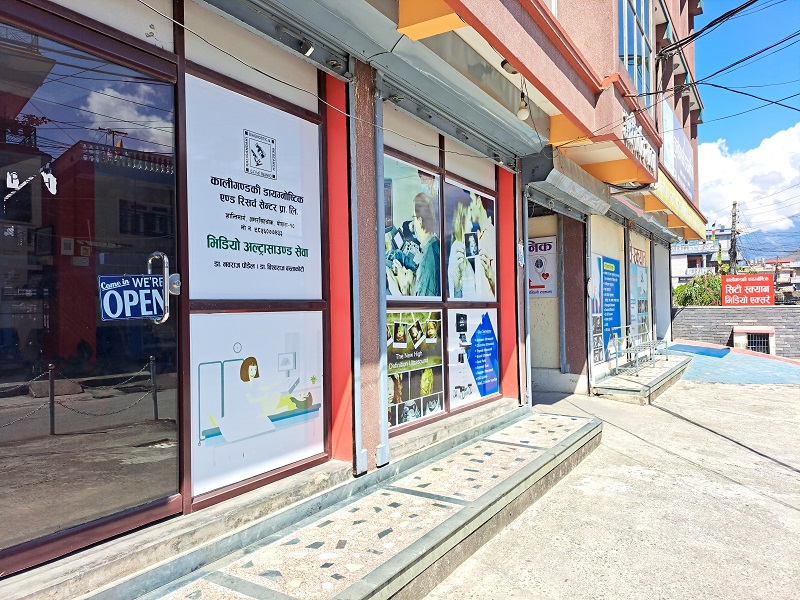
KDRC is on Shantipath at Amarsingh Chowk, Pokhara-10, Kaski. Bring your referral, earlier scans and reports, and your medicine list, and follow any fasting advice. Referring doctors can reach our team by phone or at kaligandaki.dc@gmail.com.
- Fine needle cell sampling
- Maps blood vessels before sampling
- Shows deeper lesions and wider spread
- Finds the lump and guides the needle
- Biopsies from the oesophagus and stomach
How to prepare
- Tell us about blood thinners and herbal supplements; do not stop any medicine without your prescribing doctor's advice.
- Follow any fasting instructions our staff give you; ask about diabetes medicines.
- Mention allergies (including to local anaesthetic) and pregnancy.
- Bring your referral, earlier scans, FNAC or biopsy reports.
- Bring an adult companion if sedation is planned.
- Wear comfortable clothing that gives easy access to the area.
How long it takes
Most ultrasound-guided needle biopsies are completed within about an hour, including preparation and a short period of observation.
Price in Pokhara
Biopsy cost depends on the type of biopsy, the body part, ultrasound guidance and laboratory tests required. Call 061-553538 or 9856077433 for the current price.
What to expect at KDRC
Arrive at KDRC on Shantipath, Amarsingh Chowk, with your referral, the scan report showing the area to be sampled and a list of your medicines, having followed any fasting advice. At reception, staff confirm the site, your allergies and any blood thinners. The consultant radiologist explains the biopsy, its benefits and risks, and asks for your written consent. You lie comfortably while the radiologist scans the area on HD ultrasound, often checking blood vessels with colour Doppler, and plans a safe path. The skin is cleaned and numbed with local anaesthetic, which stings briefly. Through a tiny nick, the biopsy needle takes several tissue cores under live ultrasound view; you may hear a click and feel pressure. Pressure is applied and a small dressing placed, usually without stitches. You rest briefly under observation and receive aftercare advice before going home. The samples go to a pathology laboratory, and our team tells you when to collect the report for your doctor.
Frequently asked questions
Where can I get a biopsy test in Pokhara?
Ultrasound-guided needle biopsies are available at Kaligandaki Diagnostic and Research Center (KDRC), Shantipath, Amarsingh Chowk, Pokhara-10, performed by consultant radiologists. Samples are reported by a pathologist. Call 061-553538 or 9856077433 to book, and bring your referral and the scan showing the area.
How much does a biopsy cost in Pokhara?
Biopsy cost depends on the type of biopsy, the body part, whether ultrasound guidance is needed and which laboratory tests are performed. KDRC quotes each case individually rather than publishing a fixed price. Call 061-553538 or 9856077433 for the current biopsy price before your visit.
What is a trucut biopsy?
Trucut biopsy is a common name in Nepal for a core needle biopsy. A spring-loaded hollow needle removes thin cylinders of tissue, usually under ultrasound guidance and local anaesthetic. The tissue keeps its structure, so the pathologist can make a more detailed diagnosis than from loose cells alone.
What is the difference between FNAC and biopsy?
FNAC draws loose cells through a very fine needle and is examined as cytology. A core biopsy removes small cylinders of tissue examined as histopathology, showing how cells are arranged. FNAC is quicker and gentler; a core biopsy gives more detail. Your doctor chooses the test for your situation.
Is a biopsy painful?
A needle biopsy is usually not painful because local anaesthetic numbs the area. You feel a brief sting from the injection, then pressure, and may hear a click as samples are taken. The site may be sore or bruised for a few days. Tell the team at once if you feel sharp pain.
How long does an ultrasound-guided biopsy take?
Most image-guided needle biopsies are completed within about an hour, including preparation, and the sampling itself is usually much shorter. You then rest briefly under observation before going home. Our staff will tell you how long to plan for at the centre on the day of your biopsy.
Can I eat before a biopsy?
For many needle biopsies you can eat normally. Some guided or sedated procedures need a period of fasting, sometimes around eight hours. Follow the instructions our staff give you when you book, and ask about diabetes medicines if you are asked to fast. Always mention blood thinners.
When will I get my biopsy results?
Biopsy tissue must be processed, stained and examined by a pathologist, so results usually take from a few days to a few weeks, depending on the tests needed. Our team will tell you when to collect the report. Take it to your referring doctor, who will explain the result.
Does a biopsy mean I have cancer?
No. A biopsy is done to find out what an abnormal area is, and many biopsies show benign conditions such as cysts, fibroadenomas, infection or inflammation. Only the pathology report can give the answer, so try not to assume the worst while you wait and discuss the result with your doctor.
Can a biopsy spread cancer?
Needle biopsy is a routine, well-established way to diagnose lumps, including suspected cancers, and doctors recommend it because an accurate diagnosis is essential for the right treatment. If you are worried about this, discuss it with your doctor or our radiologist before the procedure so your questions are answered.
Is a breast biopsy available in Pokhara?
Yes. KDRC offers ultrasound-guided breast biopsy, usually a core needle biopsy taking several samples under local anaesthetic. It is typically completed within an hour, no stitches are needed, and serious complications are uncommon. Ask our team whether a marker clip is planned in your case.
Can I have a biopsy during pregnancy?
Ultrasound-guided biopsy uses sound waves rather than radiation and small amounts of local anaesthetic, so it can usually be done during pregnancy when your doctor decides the result cannot wait. Always tell our staff if you are or may be pregnant so the procedure can be planned safely.
Can children have a biopsy at KDRC?
Biopsies in children are less common and need careful planning, sometimes with sedation. Please call our team with the child's referral and scan before booking. We will advise whether the biopsy can be done at KDRC or whether a paediatric centre is more suitable for your child.
What should I avoid after a biopsy?
Avoid strenuous exercise and heavy lifting for about 24 hours, keep the dressing clean and dry for the first day, and do not restart paused blood thinners until your doctor says so. Seek help for worsening pain, fever, ongoing bleeding or increasing swelling or redness at the site.
Do I need a referral for a biopsy?
Yes, in most cases. A biopsy is planned from your doctor's question and your imaging, so bring the referral slip and the ultrasound, CT or mammogram report that shows the area. If you are unsure, call 061-553538 or 9856077433 and our team will guide you on the next step.
Sources
- nhs.uk/tests-and-treatments/biopsy/
- radiologyinfo.org/en/info/biopgen
- radiologyinfo.org/en/info/breastbius
- medlineplus.gov/ency/article/003933.htm
- medlineplus.gov/ency/article/003899.htm
This page is general health information reviewed by our radiologists. It is not a diagnosis — always follow your own doctor's advice. In an emergency, call our 24-hour ambulance or go to the nearest emergency department.
Written by
Dr. Nawaraj PaudelConsultant Radiologist
Consultant Radiologist at KDRC, Pokhara. Residency at East Avenue Medical Centre, Manila; formerly Assistant Professor of Radiology at Universal College of Medical Sciences. Member, Nepal Radiologist Society.
View doctor's profile →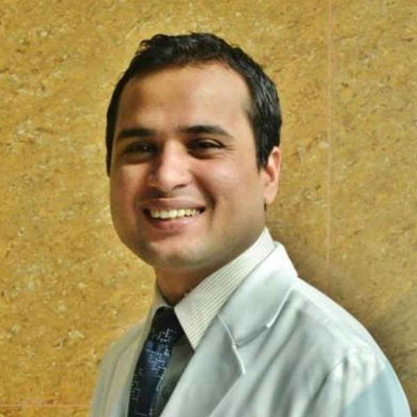
Medically reviewed by
Dr. Bishworaj BastakotiConsultant Radiologist
Consultant Radiologist at KDRC, Pokhara. Residency at the National Kidney and Transplant Institute, Manila; previously at Western Regional Hospital. Member, Nepal Radiologist Society.
View doctor's profile →Shantipath, Amarsingh Chowk, Pokhara-10, Kaski · 061553538 / 9856077433
Related services in Pokhara
192 Slice High Resolution CT Scan
A CT scan in Pokhara at KDRC is a fast, painless X-ray test that uses a computer to create detailed cross-sectional imag…
High Definition Ultrasound
Ultrasound (USG, sonography) in Pokhara at KDRC is a painless, radiation-free scan that uses high-frequency sound waves …
High Definition Endoscopy
Endoscopy in Pokhara at KDRC is an upper GI examination (gastroscopy) in which a specialist passes a thin, flexible HD c…
FNAC
An FNAC test in Pokhara at KDRC is fine needle aspiration cytology: a very thin needle draws cells from a lump, often un…
Doppler Ultrasound
A Doppler test in Pokhara at KDRC is a colour Doppler ultrasound scan that measures the speed and direction of blood flo…