
Ultrasound (USG) in Pokhara — HD 3D/4D Sonography at KDRC
Ultrasound (USG, sonography) in Pokhara at KDRC is a painless, radiation-free scan that uses high-frequency sound waves to show live images of organs, blood vessels and the unborn baby. Our HD system adds 3D, 4D and volume scanning, and every study is reported by a consultant radiologist.
Also called: USG, sonography, 4d ultrasound, 3d ultrasound, ultrasound scan, video xray
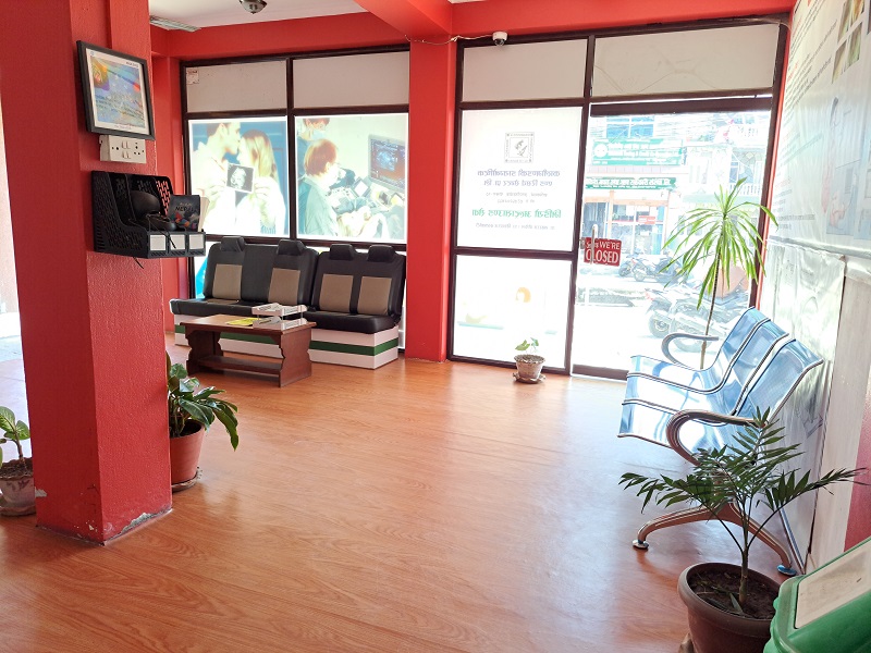
- Duration
- Most ultrasound scans take about 10–45 minutes
- Reported by
- Consultant radiologists
- Location
- Amarsingh Chowk, Pokhara-10
- Book / price
- 061553538 · 9856077433
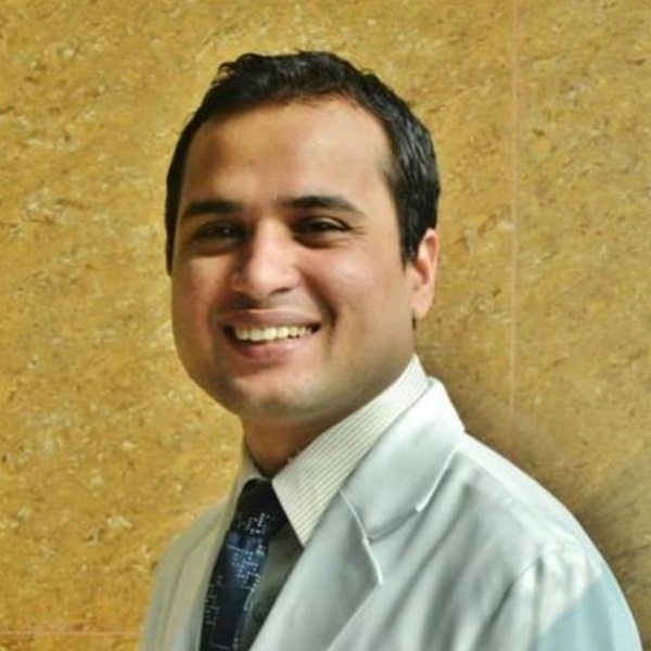 Dr. Bishworaj Bastakoti · Consultant RadiologistMedically reviewed by Dr. Nawaraj Paudel · MD Radiology Last reviewed 12 Oct 2026Updated 12 Oct 2026
- USG, sonography and “video X-ray” all mean the same radiation-free ultrasound test.
- KDRC performs abdomen, KUB, pregnancy, NT, 3D/4D, thyroid, breast, scrotal, MSK and transvaginal scans.
- Abdomen scans usually need about six hours of fasting; pelvic and early pregnancy scans need a full bladder.
- Every scan is reported by consultant radiologist Dr. Bishworaj Bastakoti or Dr. Nawaraj Paudel.
- Anomaly scans and other services are available any day of the week — call 061-553538 to book.
On this page (13 sections)
- What is an ultrasound (USG) scan?
- How does an ultrasound scan work?
- Types of USG scans available in Pokhara at KDRC
- Pregnancy ultrasound in Pokhara: which scan, which week?
- 3D and 4D ultrasound in Pokhara
- How to prepare for an ultrasound scan
- What happens during a USG scan at KDRC?
- Is ultrasound safe? Pregnancy, children and older adults
- Ultrasound vs CT vs MRI: which test is right?
- Ultrasound results and reports
- How much does an ultrasound (USG) cost in Pokhara?
- Why choose KDRC for ultrasound in Pokhara?
- Booking a USG near you in Pokhara: location and what to bring
What is an ultrasound (USG) scan?
An ultrasound scan — called USG, sonography or, in everyday Nepali, “video X-ray” (भिडियो एक्स-रे) — is a medical imaging test that uses high-frequency sound waves to create live pictures of the inside of the body. If you are looking for ultrasound in Pokhara, Kaligandaki Diagnostic and Research Center (KDRC) at Shantipath, Amarsingh Chowk, performs every common USG examination on a high-definition system with 3D, 4D and volume scanning, and each study is reported by a consultant radiologist.
Despite the popular name “video X-ray”, an ultrasound uses no X-rays and no ionising radiation at all. A small handheld probe sends sound pulses into the body and listens for the echoes. A computer turns those echoes into moving images on a screen, so the radiologist can watch organs, blood flow and an unborn baby in real time. Because it is safe, painless and quick, ultrasound is usually the first imaging test doctors in Pokhara request for tummy pain, pregnancy check-ups, lumps and swellings.
How does an ultrasound scan work?
Ultrasound works by sending sound waves that are far too high for the human ear to hear into the body and measuring how they bounce back. Different tissues reflect sound differently. Fluid, such as urine in the bladder or the amniotic fluid around a baby, lets sound pass through and looks black. Solid organs like the liver look grey. Stones and bone reflect almost all the sound and look bright white, often with a dark “shadow” behind them.
A water-based gel is spread on the skin so that no air sits between the probe and the body, because air blocks ultrasound. The same physics explains the test’s main limitation: gas in the bowel and air in the lungs scatter the sound waves, and larger body size weakens them, so some areas are harder to see clearly. RadiologyInfo notes both points. When the radiologist needs a clearer picture, your doctor may add a CT scan or MRI.
Modern machines can also measure movement. Colour Doppler shows the speed and direction of blood flow in red and blue, which helps detect clots, narrowed arteries and abnormal blood supply to a lump. Doppler is a specialised study with its own preparation and reporting, so we explain it in depth on our colour Doppler ultrasound page.
Types of USG scans available in Pokhara at KDRC
KDRC offers all routine types of ultrasound in Pokhara, from abdomen and pelvis scans to pregnancy, thyroid, breast, scrotal and musculoskeletal studies. The table below shows what each type looks at and why doctors usually ask for it.
| Type of USG | What it examines | Common reasons it is requested |
|---|---|---|
| Abdomen USG (whole abdomen) | Liver, gallbladder, bile ducts, pancreas, spleen, kidneys, aorta | Upper tummy pain, jaundice, gallstones, fatty liver, abnormal liver tests |
| Abdomen and pelvis USG | All of the above plus bladder, uterus and ovaries or prostate | General abdominal pain, swelling, check-up of pelvic organs |
| KUB ultrasound | Kidneys, ureters and urinary bladder | Kidney stones, burning urine, blood in urine, kidney function problems |
| Obstetric USG (pregnancy) | Baby, placenta, amniotic fluid, cervix | Confirming and dating pregnancy, growth, position, wellbeing |
| NT scan | Fluid at the back of the baby’s neck, early anatomy | First-trimester screening at about 11–14 weeks |
| 3D / 4D ultrasound | Surface and volume views, mostly of the baby | Clearer views of the face, limbs and selected structures |
| Transvaginal scan (TVS) | Uterus, ovaries, early pregnancy | Early pregnancy, irregular bleeding, ovarian cysts, fertility checks |
| Thyroid and neck USG | Thyroid gland, lymph nodes, salivary glands | Neck swelling, thyroid nodules, enlarged glands |
| Breast ultrasound | Breast tissue and armpit lymph nodes | Breast lump, pain, nipple discharge, follow-up of a known lump |
| Scrotal ultrasound | Testes, epididymis, scrotal contents | Pain, swelling, lump, hydrocele, varicocele |
| Musculoskeletal USG | Tendons, muscles, joints, soft-tissue lumps | Sports injuries, shoulder pain, swellings, fluid collections |
Abdomen, pelvis and KUB ultrasound
Abdomen USG is the most requested scan in Pokhara. It shows gallstones, fatty liver, liver lumps, swollen bile ducts, kidney stones, kidney swelling (hydronephrosis) and fluid in the abdomen. A KUB scan focuses on the urinary tract and is the usual first test for flank pain and urine symptoms. In women a pelvic scan checks the uterus and ovaries for fibroids and cysts; in men it measures the prostate and checks how well the bladder empties.
Pregnancy (obstetric) ultrasound
Pregnancy ultrasound in Pokhara follows the same schedule used internationally: an early dating scan, an NT scan at about 11–14 weeks, the detailed anomaly scan in mid-pregnancy, and growth scans later when needed. The detailed mid-pregnancy check has its own page — read about our anomaly scan in Pokhara (Level II ultrasound), which is available any day of the week at KDRC.
Thyroid, breast and scrotal ultrasound
Small-parts ultrasound uses a high-frequency probe that shows fine detail close to the skin. It tells whether a lump is a simple fluid-filled cyst or a solid nodule, measures it, and checks its blood supply. If a nodule needs a sample, the radiologist can guide a fine needle into it under ultrasound — see FNAC and ultrasound-guided biopsy.
Musculoskeletal and soft-tissue ultrasound
Ultrasound can show tendons and muscles while you move, which is something static scans cannot do. It is useful for shoulder pain, tendon tears, Baker’s cysts behind the knee, abscesses and lumps under the skin. Deep joint and bone problems are usually better shown by MRI.
Ultrasound-guided procedures
Because ultrasound shows a needle moving in real time, it is the safest way to guide many minor procedures without radiation. At KDRC the radiologist uses ultrasound guidance to sample thyroid nodules, lymph nodes and breast lumps, and to place a needle or tube accurately when fluid needs to be drained. Guidance lets the radiologist avoid blood vessels and reach small targets with a single, precise pass. The details of each procedure — consent, preparation and aftercare — are explained on the FNAC, biopsy and catheter insertion pages, so the ultrasound itself simply becomes the radiologist’s eyes during the procedure.
Transvaginal (TVS) ultrasound
A transvaginal scan uses a slim, covered probe placed gently in the vagina, close to the uterus and ovaries. The NHS notes that internal probes can be uncomfortable but that you can ask the operator to stop at any time. A female staff member or chaperone can be present, and the scan is only done with your consent.
| Feature | Transabdominal (over the tummy) | Transvaginal (TVS) |
|---|---|---|
| Probe position | On the skin of the lower abdomen | Slim covered probe inside the vagina |
| Bladder | Usually full | Empty |
| Best for | General pelvic view, later pregnancy | Early pregnancy, ovaries, uterine lining |
| Image detail | Good overview | Higher detail of nearby organs |
| Comfort | Painless; pressure from full bladder | May be mildly uncomfortable; you can ask to stop |
Pregnancy ultrasound in Pokhara: which scan, which week?
Most pregnancies need at least two key ultrasound scans: a dating or NT scan at 11–14 weeks and an anomaly scan at 18–21 weeks, according to the NHS. Your obstetrician may add early or growth scans depending on your health and pregnancy. The table summarises the usual timing.
| Scan | Usual timing | Main purpose |
|---|---|---|
| Early pregnancy scan | First weeks after a positive test, if your doctor requests it | Confirms the pregnancy is in the uterus, heartbeat, number of babies |
| Dating and NT scan | About 11–14 weeks | Due date, number of babies, NT measurement for screening |
| Anomaly scan (Level II, TIFFA) | About 18–21 weeks (often described as 18–22) | Detailed organ-by-organ check of the baby, placenta and fluid |
| Growth scan | Third trimester, when your doctor needs it | Baby’s size, fluid volume, position and wellbeing |
The NT measurement is taken during the dating scan and is combined with a blood test for Down’s syndrome screening; the NHS notes that this combined test must be done before the end of the 14th week. If you live outside Pokhara or cannot travel late in pregnancy, ask about our mobile (home) ultrasound service.
3D and 4D ultrasound in Pokhara
A 3D ultrasound builds a three-dimensional image from many thin slices, and a 4D ultrasound is simply 3D shown in motion, as Cleveland Clinic explains. KDRC’s HD system also records volume data, so the radiologist can review the scanned area from different angles after you leave the couch.
In pregnancy, 3D and 4D views can make the baby’s face, lips and limbs easier to understand, and many families value seeing them. They do not replace the standard 2D measurements, which remain the basis of the medical report. The US FDA discourages ultrasound done purely for keepsake videos, but accepts keepsake images taken during a medically needed scan when no extra exposure is added. That is how we offer 3D/4D at KDRC — as part of a clinically requested examination, often the anomaly scan or a growth scan.
How to prepare for an ultrasound scan
Preparation for an ultrasound depends on the area being scanned: abdominal scans need an empty stomach, while pelvic and early pregnancy scans need a full bladder. Our staff confirm the exact instructions when you book on 061-553538 or 9856077433.
| Scan type | Preparation | What to bring or wear |
|---|---|---|
| Abdomen USG | No food for about 6 hours; small sips of water are usually fine | Doctor’s request slip, old reports |
| Abdomen and pelvis / KUB | Fast as above, then drink water and do not empty your bladder | Loose two-piece clothing |
| Pregnancy scan (early) | Usually a comfortably full bladder | Pregnancy card, previous scan reports |
| Pregnancy scan (later) | Usually no special preparation | Antenatal card, previous scans and blood reports |
| Transvaginal scan | Empty your bladder just before the scan | A companion or chaperone if you wish |
| Thyroid, breast, scrotal, MSK | No preparation | Clothing that is easy to remove from the area; any earlier mammogram or reports |
The six-hour fasting guide for abdominal scans matches common hospital practice — for example Whittington Health NHS asks for six hours without food — because fasting keeps the gallbladder full and reduces bowel gas. Keep taking your regular medicines with a little water unless your doctor says otherwise, and tell us if you have diabetes so we can schedule your fasting scan early.
- Ask us which preparation your scan needs when you book
- Abdomen scan: no food for about 6 hours; sips of water are fine
- Pelvic or early pregnancy scan: arrive with a comfortably full bladder
- Bring your doctor's request slip and previous reports
- Bring your antenatal card if you are pregnant
- Wear loose two-piece clothing
- Take regular medicines unless your doctor says otherwise
What happens during a USG scan at KDRC?
During a USG scan you lie on a couch while the operator moves a gel-covered probe over your skin and watches live images on a screen. Most patients find it relaxing; you may feel light pressure when the probe presses to look past gas. The NHS gives a typical range of about 10 to 45 minutes depending on the examination.
- 1Book and prepareCall 061-553538 or 9856077433, confirm the scan type and follow the fasting or full-bladder instructions.
- 2Register at receptionShow your request slip and previous reports at our Shantipath reception.
- 3Get positionedLie on the couch and uncover only the area to be scanned; a chaperone can be present.
- 4Gel and probeWarm gel is applied and the probe is moved over the skin while live images appear on screen.
- 5Images reviewedThe radiologist reviews the images and may ask you to hold your breath or turn.
- 6Collect your reportWipe off the gel, resume normal activity, and collect the report when our team tells you it is ready.
Is ultrasound safe? Pregnancy, children and older adults
Yes — diagnostic ultrasound is considered safe because it uses sound waves, not ionising radiation, and has no known harmful effects at the energy levels used for scanning (RadiologyInfo). The NHS states there are no known risks to the baby or the mother from a pregnancy scan.
Ultrasound energy can slightly warm tissue, so responsible centres follow the ALARA principle — “as low as reasonably achievable” — using only the energy and time needed for a diagnostic image, as recommended by the FDA. Our radiologists apply ALARA to every scan, especially in pregnancy.
- Pregnancy: ultrasound is the standard imaging test in pregnancy and can be repeated when medically needed.
- Children and babies: no radiation and no needles, so it is ideal for tummy pain, kidney problems and lumps in children. A parent stays in the room.
- Older adults and frail patients: the scan is done lying comfortably, and a home visit is possible through mobile ultrasound.
- Implants and pacemakers: ultrasound is not affected by metal, so patients who cannot have MRI can still be scanned.
Ultrasound vs CT vs MRI: which test is right?
Ultrasound is usually the first choice when a radiation-free, quick and widely available test can answer the question; CT and MRI are used when deeper or more detailed images are needed. Your doctor decides which test fits your symptoms. The comparison below shows the general differences.
| Feature | Ultrasound (USG) | CT scan | MRI |
|---|---|---|---|
| Uses | Sound waves | X-rays | Magnetic field and radio waves |
| Ionising radiation | None | Yes | None |
| Live, moving images | Yes | No | Limited |
| Best for | Pregnancy, gallbladder, kidneys, thyroid, breast, scrotum, soft tissue | Trauma, lungs, stones, bleeding, bones, whole-body staging | Brain, spine, joints, soft-tissue detail |
| Limited by | Bowel gas, bone, larger body size | Radiation; contrast in kidney disease | Some implants; lying still for longer |
| Safe in pregnancy | Yes, standard test | Only if clearly needed | Used selectively |
All three tests are available in one building at KDRC — our 192-slice CT scan and open-type MRI pages explain when each is used.
Ultrasound results and reports
Your ultrasound is interpreted by a consultant radiologist, who writes a report for your referring doctor. Our team will tell you when to collect your report. The report describes the size and appearance of each organ, measurements of anything abnormal, and the radiologist’s impression.
Please take the report to the doctor who requested the scan. Only your doctor can combine the result with your symptoms, examination and blood tests to make a diagnosis and plan treatment. Bring earlier reports so changes over time can be compared. Referring hospitals and clinics can discuss urgent findings with the radiologist, and images can be shared through our teleradiology service.
Go to an emergency department straight away for severe abdominal pain, heavy vaginal bleeding or severe pain in pregnancy, fainting, sudden severe testicular pain, or a pregnant woman who notices the baby moving much less. KDRC's ambulance can help with transport — call 061-553538 or 9856077433.
How much does an ultrasound (USG) cost in Pokhara?
The cost of an ultrasound in Pokhara depends on the type of scan, so KDRC quotes each examination individually rather than publishing a fixed price list. A single-organ scan, a whole abdomen and pelvis study, a detailed pregnancy scan and a 3D/4D add-on each take different time and expertise. Adding Doppler, ultrasound guidance for a needle procedure, or a home visit also changes the cost. For the current USG price in Pokhara, call 061-553538 or 9856077433 (Viber) — our staff will tell you the price and the preparation at the same time.
Why choose KDRC for ultrasound in Pokhara?
Patients and doctors choose KDRC for ultrasound in Pokhara because every scan is done on a high-definition 3D/4D system and reported by an experienced consultant radiologist, with CT, MRI and Doppler available under the same roof.
- HD ultrasound with 3D, 4D and volume scanning, plus colour Doppler in the same session when your doctor requests it.
- Consultant radiologists: Dr. Bishworaj Bastakoti (residency at the National Kidney and Transplant Institute, Manila; Diplomate, Philippine College of Radiology) and Dr. Nawaraj Paudel (residency at East Avenue Medical Centre, Manila; former assistant professor of radiology). Both are members of the Nepal Radiologist Society and NMC-registered radiologists since 2016. Meet them on our doctors page.
- Pregnancy scans any day of the week, including the detailed anomaly scan.
- One-stop diagnostics: if your scan needs follow-up, FNAC, biopsy, CT or MRI can be arranged in the same centre.
- Home and bedside scanning through mobile ultrasound, and an ambulance service for patients who need transport.

Booking a USG near you in Pokhara: location and what to bring
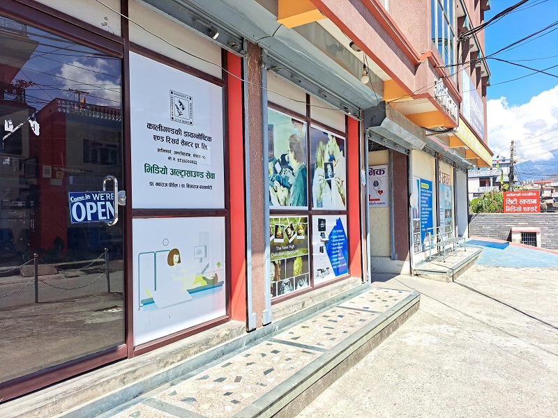
KDRC is at Shantipath, Amarsingh Chowk, Pokhara-10, Kaski — easy to reach by taxi or local bus from Lakeside, Prithvi Chowk, Mahendrapul and Bagar. If you searched “USG near me” from anywhere in Pokhara, call 061-553538 or 9856077433 (also on Viber) to book a time, or use our online appointment form. We can give you the preparation instructions on the same call.
Bring your doctor’s request slip (a referral is helpful but not always required — ask us), any previous ultrasound, CT or MRI reports, your antenatal card if pregnant, and a list of your medicines. Wear loose two-piece clothing. If you need a different kind of imaging, browse all KDRC services.
- detailed 18–22 week pregnancy scan
- home and bedside scanning
- ultrasound-guided tissue sample
- blood-flow study
- ultrasound-guided needle sample
- when more detail is needed
How to prepare
- Call 061-553538 or 9856077433 to confirm the preparation for your scan type.
- Abdomen USG: no food for about 6 hours; small sips of water are fine.
- Abdomen and pelvis or KUB: fast, then drink water and keep your bladder full.
- Early pregnancy or pelvic scan: arrive with a comfortably full bladder.
- Transvaginal scan: empty your bladder just before the scan.
- Thyroid, breast, scrotal and musculoskeletal scans: no preparation.
- Bring your request slip, previous reports and antenatal card; wear loose two-piece clothing.
How long it takes
Most ultrasound scans take about 10–45 minutes, depending on the area examined and how clearly it can be seen.
Price in Pokhara
USG cost in Pokhara depends on the scan type, the number of areas examined and add-ons such as 3D/4D, Doppler or a home visit. Call 061-553538 or 9856077433 for the current price.
What to expect at KDRC
When you arrive at KDRC on Shantipath, Amarsingh Chowk, register at reception with your doctor's request slip, previous reports and, if pregnant, your antenatal card. Staff check that you have followed the preparation — fasting for an abdomen USG, a full bladder for a pelvic or early pregnancy scan — and tell you roughly how long you will wait. In the scan room you lie on a couch and uncover only the area being examined; a family member or chaperone can stay with you. Warm gel is spread on the skin and the probe glides over it while live images appear on the screen. You may be asked to take a deep breath, hold it, or turn onto your side so organs move out from behind bowel gas. Most scans take between ten and forty-five minutes. If 3D/4D views are part of a pregnancy scan, they are captured during the same session. Afterwards you wipe off the gel, empty your bladder if needed, and can eat, drive and return to work straight away. Our team tells you when to collect your report. Take it to the doctor who requested the test.
Frequently asked questions
Where can I get an ultrasound (USG) in Pokhara?
You can get an ultrasound at Kaligandaki Diagnostic and Research Center (KDRC), Shantipath, Amarsingh Chowk, Pokhara-10. We perform abdomen, pelvis, KUB, pregnancy, 3D/4D, thyroid, breast, scrotal, musculoskeletal and transvaginal scans on an HD system, reported by consultant radiologists. Call 061-553538 or 9856077433 to book a time.
What is the USG price in Pokhara?
The USG price depends on the type of scan — a single organ, whole abdomen and pelvis, detailed pregnancy scan, 3D/4D add-on or Doppler each differ. KDRC quotes prices individually and updates them from time to time, so please call 061-553538 or 9856077433 (Viber) for the current price before your visit.
Is a video X-ray the same as an ultrasound?
Yes. In Nepal, people often call ultrasound “video X-ray” (भिडियो एक्स-रे) because it shows moving pictures. However, it uses no X-rays at all. Ultrasound creates images from sound waves, so it carries no radiation and is safe for pregnant women and children.
Do I need to fast before an ultrasound?
Only for some scans. An abdomen USG usually needs about six hours without food so the gallbladder is full and bowel gas is reduced; small sips of water are usually fine. Thyroid, breast, scrotal and later pregnancy scans need no fasting. Our staff confirm your preparation when you book.
Why do I need a full bladder for a pelvic or pregnancy scan?
A full bladder pushes bowel gas out of the way and acts as a clear window for sound waves to reach the uterus, ovaries, prostate or early pregnancy. Drink water before the scan and avoid emptying your bladder until it is done. For a transvaginal scan, the bladder should be empty instead.
Is ultrasound safe during pregnancy?
Yes. Ultrasound uses sound waves, not radiation, and the NHS states there are no known risks to mother or baby from a pregnancy scan. At KDRC our radiologists follow the ALARA principle, using only the energy and scan time needed for a clear diagnostic image.
How long does an ultrasound take?
Most ultrasound scans take about 10 to 45 minutes, depending on the area examined and how clearly it can be seen. A thyroid or single-organ scan is usually quick, while a detailed pregnancy scan or a combined abdomen and pelvis study takes longer. Allow extra time for registration on the day.
Is ultrasound painful?
No. An external ultrasound is painless; you feel cool gel and light pressure from the probe. If the area is already tender, pressing may be briefly uncomfortable. A transvaginal scan can feel mildly uncomfortable, but it should not hurt, and you can ask the operator to stop at any time.
Can children have an ultrasound?
Yes. Ultrasound is the preferred first test for many problems in children because it uses no radiation, no needles and usually no sedation. A parent stays with the child throughout. Bring a favourite toy or snack for after the scan, and tell us when booking if your child needs to fast.
What is the difference between 3D and 4D ultrasound?
A 3D ultrasound creates a still three-dimensional image from many thin slices, while 4D ultrasound shows that 3D image moving in real time. At KDRC, 3D/4D views are added to a medically requested scan — usually a pregnancy scan — to show the baby's face and limbs more clearly.
When will I get my ultrasound report?
Ultrasound reports for routine scans are often ready soon after the scan, and complex studies may take longer. Our team will tell you when to collect your report. Take it to the doctor who requested the scan, who will explain what the findings mean for you.
Do I need a doctor's referral for an ultrasound in Pokhara?
A doctor's request is recommended because it tells the radiologist what question to answer and ensures someone will act on the result. If you do not have one, call us on 061-553538 or 9856077433 — our staff will advise you, and for pregnancy scans please bring your antenatal card.
Is ultrasound available on Saturdays at KDRC?
KDRC offers anomaly scans and other services any day of the week. Because scheduling can vary, please call 061-553538 or 9856077433 before coming on a Saturday or public holiday so we can confirm a time and give you the right preparation.
Can ultrasound detect cancer?
Ultrasound can show lumps and tell whether they are fluid-filled cysts or solid, and it can reveal features that need further tests. It cannot confirm cancer on its own. If needed, a sample can be taken under ultrasound guidance through FNAC or biopsy, or your doctor may request CT or MRI.
Can I get an ultrasound at home in Pokhara?
Yes. KDRC's mobile ultrasound service brings a portable machine to patients who cannot easily travel, such as elderly, bedridden or post-operative patients. The images are reported by our consultant radiologists in the same way. Call 061-553538 or 9856077433 to arrange a home scan.
Sources
- nhs.uk/tests-and-treatments/ultrasound-scan/
- radiologyinfo.org/en/info/genus
- radiologyinfo.org/en/info/abdominus
- nhs.uk/pregnancy/your-pregnancy-care/ultrasound-scans/
- nhs.uk/pregnancy/your-pregnancy-care/12-week-scan/
- fda.gov/radiation-emitting-products/medical-imaging/ultrasound-imaging
- my.clevelandclinic.org/health/diagnostics/4995-ultrasound
- whittington.nhs.uk/?c=44313
This page is general health information reviewed by our radiologists. It is not a diagnosis — always follow your own doctor's advice. In an emergency, call our 24-hour ambulance or go to the nearest emergency department.
Written by
Dr. Bishworaj BastakotiConsultant Radiologist
Consultant Radiologist at KDRC, Pokhara. Residency at the National Kidney and Transplant Institute, Manila; previously at Western Regional Hospital. Member, Nepal Radiologist Society.
View doctor's profile →Medically reviewed by
Dr. Nawaraj PaudelConsultant Radiologist
Consultant Radiologist at KDRC, Pokhara. Residency at East Avenue Medical Centre, Manila; formerly Assistant Professor of Radiology at Universal College of Medical Sciences. Member, Nepal Radiologist Society.
View doctor's profile →Shantipath, Amarsingh Chowk, Pokhara-10, Kaski · 061553538 / 9856077433
Related services in Pokhara
Anomaly Scan (Level II Ultrasound)
An anomaly scan in Pokhara at KDRC is a detailed Level II (TIFFA) pregnancy ultrasound, usually done at 18–22 weeks, tha…
FNAC
An FNAC test in Pokhara at KDRC is fine needle aspiration cytology: a very thin needle draws cells from a lump, often un…
Biopsy
A biopsy test in Pokhara at KDRC is a procedure in which a small tissue sample is taken from a lump or abnormal area, us…
Doppler Ultrasound
A Doppler test in Pokhara at KDRC is a colour Doppler ultrasound scan that measures the speed and direction of blood flo…
Mobile Ultrasound
Mobile ultrasound service in Pokhara at KDRC brings a portable ultrasound machine and trained team to the bedside of pat…